|
Test whether the process degrades the images and if they contain all the diagnostic information possible
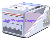
A multi format imager device is currently the most common method for presenting the imaging information to the diagnostician and for archiving it.
Multi format imagers may be used to present imaging information from the gamma camera and archive the results of computer processing.
-
Multi format imager used to imprint images of a gamma camera must be examined daily to be sure that:
-
Its dot sharpness and size, intensity, and image shape are correct. This is accomplished as part of the daily uniformity testing.
A simple procedure may be:
-
Place the Low Energy High Resolution (LEHP) collimator on the camera. Turn the camera face vertically upward.
-
Place the Co57 flat source upon the collimator.
-
Center the 20% energy window on the radionuclide photopeak.
-
Acquire an image of enough counts and in the highest pixel matrix size.
-
Inspect the flashes on the screen. Adjust the focus and astigmatism controls so that flashes are small and circular.

A Cathode - Ray Tube (CRT) screen malfunction may cause unfocused flashes. If lines are presented instead of points the pulse arithmetic and timing circuits may need adjustment.
-
The following are also recommended to obtain optimum nuclear medicine imaging by a multi format imager :
-
Sharpness and acceptable film density are examined for each image.
-
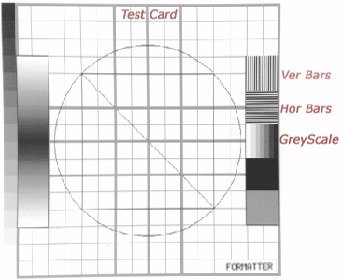
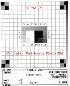
Store in the computer the image of sets of gray levels representing those that are used in the patient images.
-
Photograph this image and examine it to see how many of the gray levels are visible on the film.
-
The imager contrast, brightness and exposure time must be adjusted until the number of gray levels is acceptable.
-
If the range of the gray scale of the film is used, it is possible to obtain good quality images that should not be so light as to appear transparent nor so dark as to obscure detail in the images.
-
If the diagnostic interest in the images produced by the imager is in the low-count portion of the range, make sure that, low levels of gray can be accurately portrayed in the image.

If the multi-format imager is not drifting, the testing of the gray levels may be done weekly.
|
 Test of the film Imager